New 16-slice Computed Tomography scanner installed in Veterinary Teaching Hospital
A new Toshiba Aquilion 16-slice Computed Tomography (CT) scanner that will significantly improve imaging quality and speed has been installed in the Horace E. and Elizabeth F. Alphin Radiology Center Virginia-Maryland Regional College of Veterinary Medicine at Virginia Tech's Veterinary Teaching Hospital.
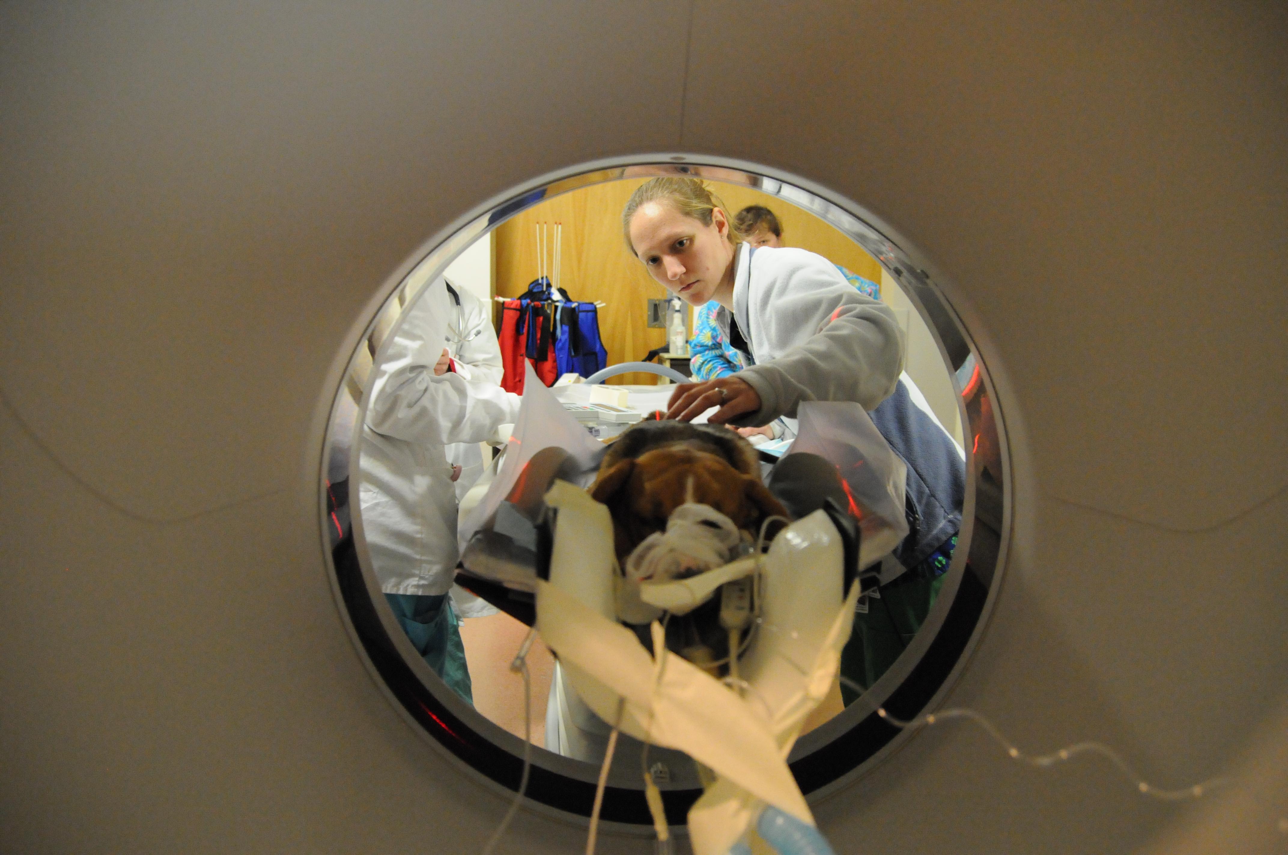
CT scans provide high-resolution cross-sectional anatomical images. Patients are placed on a table that moves through a circular opening in the scanner, called the gantry, while an x-ray tube emits rays as it spins 360 degrees. A detector array measures the energy of x-rays that pass through the anatomic area of interest and cross-sectional images are generated from this data.
With the new scanner, which is approximately five-times faster than its predecessor, the average scanning time is so rapid patients can be examined under sedation rather than general anesthesia. Moving structures such as the lungs, heart, and blood vessels can also be more accurately assessed.
“We can scan the entire chest of a dog in approximately 20 seconds with very thin slices and very high detail,” explains Dr. Jeri Jones, an associate professor and radiologist in the Department of Small Animal Clinical Sciences.
The scanner is linked to an image analysis workstation, automatic contrast injector, laser imager, and an off-site storage server. The image analysis workstation is used to create two- and 3-D images which significantly help the radiologists with their diagnosis and clinicians with their treatment plans. The automatic contrast injector enables the patients to be injected with a contrasting material to allow their blood vessels and arteries to be imaged.
The most common anatomic regions currently imaged with the hospital’s CT scanner are the spine and head. However, the scanner is also used for imaging the musculoskeletal system, abdomen, and chest. Faculty members are in the process of developing a new protocol which will enable them to image abnormalities of the heart.
“We now have the ability to look inside our patients with a non-invasive technique that is very fast, so we don’t always have to put them under anesthesia.” said Jones. “This new technology is especially helpful for determining the extent of disease involvement and planning treatments in complex anatomic regions. The ability to create three-dimensional displays also makes the CT findings more understandable for our clients so they can make more informed decisions.”