Virginia-Maryland Regional College of Veterinary Medicine acquires MRI unit
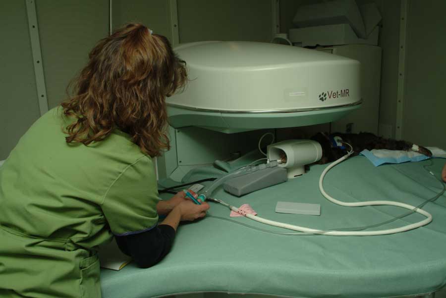
The Virginia-Maryland Regional College of Veterinary Medicine (VMRCVM) has expanded the range of non-invasive procedures available in the Veterinary Teaching Hospital (VTH) with the acquisition of a Magnetic Resonance Imaging (MRI) unit.
Unlike conventional x-rays and computed tomography (CT) which use ionizing radiation to create diagnostic images, MRI uses a strong magnetic field and radio waves to create detailed images of internal structures, according to veterinary radiologist Dr. Jeryl Jones, an associate professor in the Department of Small Animal Clinical Sciences.
MRI is considered the most sensitive diagnostic imaging test for brain and spinal cord diseases since it can detect extremely subtle abnormalities.
"We are very happy and excited to now have an in-house MRI scanner,” said Dr. Jones, who is board-certified by the American College of Veterinary Radiology. “This advanced imaging technology further increases our diagnostic capabilities and allows us to continue to provide outstanding care in a timely manner to our clients and patients. It is also a valuable teaching tool that will help us better serve the needs of our students, interns, and residents."
When placed in a MRI unit, the hydrogen atoms within tissues align with a strong magnetic field generated by the instrument, according to Dr. Jones. Radio waves pulsed into the field then stimulate the hydrogen atoms to release energy, which is transmitted to a computer for analysis, she explains. Since the signals of abnormal tissues are different from those received from normal tissues, they will show up as either very white or very dark areas in the computer image display.
The new unit was installed in December 2006. The first patient scan was performed in early January and around 20 scans have been performed since then, said to Dr. Jones.
Brain masses and spinal cord compression are the most common diagnoses made by the MRI unit. These conditions are most commonly treated through surgery or, in the case of tumors, radiation therapy.
While currently used primarily for the diagnosis and treatment of injuries and diseases of the spinal cord and brain, Dr. Jones predicts the unit will also likely be used to diagnose canine knee joint problems in the future.
The new unit is being leased with a current contract of three years. Leasing high-technology imaging equipment as opposed to purchasing allows lower up-front costs and more frequent upgrades, according to Dr. Jones. This benefits the patient, client, and clinician because it helps the VTH keep fees as low as possible while offering modern diagnostic and therapeutic technologies.
Before the installation of the new MRI unit, VMRCVM clinicians had to send patients to alternative veterinary MRI facilities, such as those in Vienna, Va., and Raleigh, N.C., both owned by the pet food company, IAMS®.
The Virginia-Maryland Regional College of Veterinary Medicine (VMRCVM) is a two-state, three-campus professional school operated by the land-grant universities of Virginia Tech in Blacksburg and the University of Maryland at College Park. Its flagship facilities, based at Virginia Tech, include the Veterinary Teaching Hospital, which treats more than 40,000 animals annually. Other campuses include the Marion duPont Scott Equine Medical Center in Leesburg, Va., and the Avrum Gudelsky Veterinary Center at College Park, home of the Center for Government and Corporate Veterinary Medicine. The VMRCVM annually enrolls approximately 500 Doctor of Veterinary Medicine and graduate students, is a leading biomedical and clinical research center, and provides professional continuing education services for veterinarians practicing throughout the two states. Virginia Tech, the most comprehensive university in Virginia, is dedicated to quality, innovation, and results to the commonwealth, the nation, and the world.
.jpg.transform/m-medium/image.jpg)